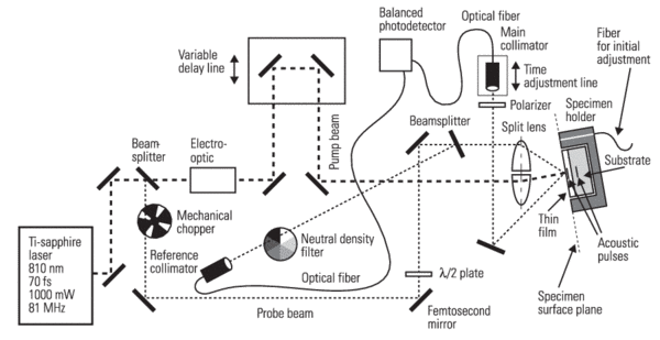
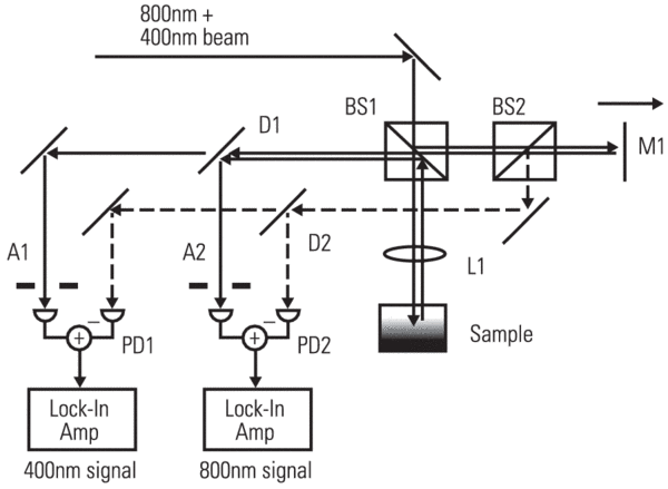
Owing to the advent of femtosecond Ti:sapphire pulsed lasers (with pulses as short as only a few optical cycles), the ultrafast pump-probe method has been successfully adapted to detect small, time-domain signals in many areas. New Focus balanced photoreceivers are particularly well suited to optical, time-resolved pump-probe measurements as in Figure 2.
One example associated with the balanced photodetection technique is femtosecond ultrasonics wherein a femtosecond laser pulse is used to excite an acoustic wave in a material. The length of mechanical (acoustic) wave determines the resolution of ultrasound. Depending upon the materials for test, the velocity of sound, propagating through the media, has a magnitude in the order of 103 m/s. The acoustic wavelength employed in classical ultrasonics locates at around 0.1–10 mm, depending on materials and frequencies. A growing demand of computer chip manufacturers for non-destructive testing of microstructures and thin films has pushed the wavelength scope down to 10–20 nm.
Piezoelectric devices used for production and echo detection of acoustic waves in the macroscopic scale are too rigid in order to resolve signals within time scales of a few picoseconds and corresponding frequencies of 0.30.6 THz. In 1987, researchers at Brown University4 proposed the use of laser-generated ultrasound for film thickness measurements. The performance of the laser-based acoustic method has been further improved recently by means of double-frequency modulation, cross-polarization, and balanced photodetection techniques5. Shown in Figure 2 is an improved pump-probe laser-based ultrasonic set-up as it is realized at the Center of Mechanics, Swiss Federal Institute of Technology in Zürich. The specimens (DUTs) consist of aluminum film (with 100, 200, or 300 nm in thickness) on a sapphire substrate.
A Ti:sapphire laser is used in this event to create short laser pulses having durations of less than 70 fs (1015 s) and a wavelength of 810 nm at a repetition rate of 81 MHz. The laser beam is split into a pump beam (carrying 90% of the energy) and a weaker probe beam by a beamsplitter. The short pump pulse hits perpendicular to the surface of the film specimen, and is absorbed within a thin surface layer (less than 10 nm deep). A mechanical stress is generated, which then excites thermo-elastically an acoustic pulse. When the bulk wave propagates and hits a discontinuity of the acoustic impedance (note: the film substrate border represents a strong discontinuity of the acoustic impedance), an echo occurs which is heading back to the surface of the film. Reaching the surface, the echo causes a slight change of the optical reflectivity.
The purpose of the probe pulse is to scan the optical reflectivity at the thin film surface versus time. Therefore, the experiments are constantly repeated at a repetition rate of 81 MHz, while the length of the optical path of the pump beam is varied. This means that the relative time shift between the pump pulse and the probe pulse is varied, and the optical reflectivity at the surface is scanned versus this relative time shift.
The change of the optical reflectivity caused by the acoustic echoes is very small, and typically well below the noise level. Improvements need to be added, in order to reduce optical and electric crosstalk between the excitation and the detection, as well as to separate the signal of interest from surrounding noises. Thus, the goal of the set-up is to isolate the thermal-acoustic phenomena occurring at the specimen as the only bridge carrying information from the excitational side (pump beam) to the measuring side (probe beam).
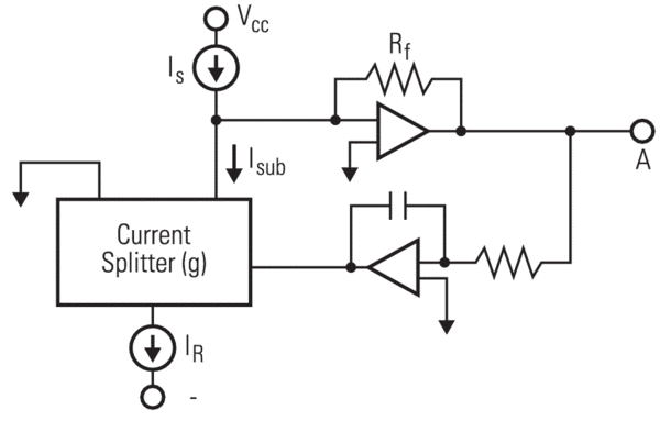
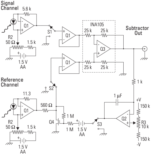
Various features are introduced to this set-up. Double-frequency modulation: The pump beam and the probe beam are modulated at different frequencies. The pump beam is modulated harmonically at a frequency of 19 MHz, and the probe beam is chopped mechanically at a frequency of 300 Hz. A dual lock-in amplification scheme is applied in order to extract the signal caused by the acoustic echoes. Cross-polarization: The pump pulses and the probe pulses propagate in different polarization planes. Therefore, a plate is shifted into the probe beam; in order to turn the original vertically polarized light into a horizontal plane. Balanced photodetection: A balanced photodetector (such as New Focus™ Model 1607) is receiving light from the two probe beams (i.e., one signal reflects at the specimen and one reference beam reflects at a beamsplitter before it hits the specimen) through two single-mode fibers. The intensities and the phase shift of these two probe pulses are carefully equalized before the difference of its intensities is measured and amplified by the balanced photodetector.
These added features show two major advantages: first, the detected signal is caused by the optical reflectivity change only, and, secondly, fluctuations of the intensity are cancelled out by the balanced photoreceiver, thus, reduce the noise level in general. The sensitivity improvement of this set-up is estimated to amount to one to two orders of magnitude. Applications need non-destructive, in-situ metrology, such as small-scale material characterization, MEMS inspection, etc., will see benefits from this femtosecond ultra-sonic method.